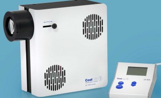
Capture high-speed events
The pE-400max offers both global and individual channel TTL triggering.
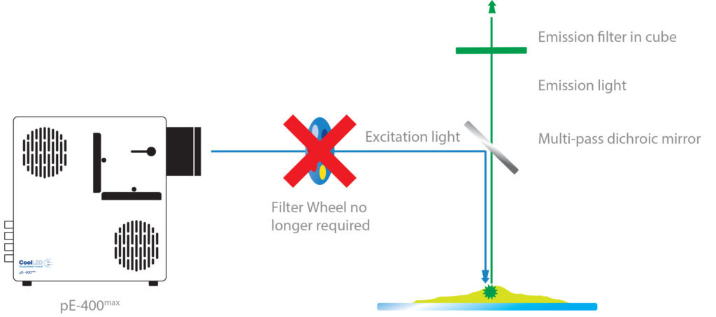
For capturing fast events in multi-stained samples, users can capitalise on high-speed <10 µs TTL triggering by utilising optional filter holders. These can accommodate single-band excitation filters in the optical path of each channel, as shown below:
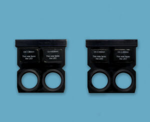
Optional filter holders sold separately
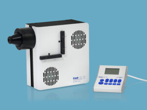
pE-400max liquid light guide with optional filter holders attached
High-speed imaging is achieved with the pE-400max thanks to <10 µs TTL triggering and optional filter holders. In this setup, individual LED channel switching is controlled via TTL triggering, and single-band excitation filters are housed in the Light Source as part of a Pinkel configuration (where multi-band dichroic and emission filters are housed in a filter cube). This overcomes the cost and latency restrictions of a filter wheel, presenting a cost-effective approach to multi-channel imaging.
For live cell imaging applications, events can now be captured that might previously have been missed. Read our white paper to compare the benefits and trade-offs of different possible illumination configurations.
Affordable automation with Sequence Runner
The popular Sequence Runner program combines the versatility of software with high-speed TTL, with minimal requirement for expensive electronic hardware. Once an LED and irradiance sequence are specified, the sequence cycles through automatically – with triggering synchronised via the global TTL-in of the pE-400max and a single TTL-out from a camera. When combined with inline filters, this transforms a manual microscope into a cost-effective four-channel automated imaging system.
Protecting your samples
Tight hardware synchronisation not only increases temporal resolution, but also means samples are exposed only during acquisition, protecting against photobleaching and phototoxicity and pushing the boundaries of time-lapse studies.
In the LightBridge graphical user interface (see below), live samples can be further protected by balancing the irradiance to the lowest level possible while still maintaining image quality, with control in 1% steps up to 100%. And the more life-like a cell behaves, the more valuable a data set becomes.
Intuitive control
For quick and simple operation, the pE-400max can be controlled with manual control pod. For more complex experiments, sophisticated control is possible with third-party imaging software (via USB Type B connection) and users can benefit from full integration into µManager.
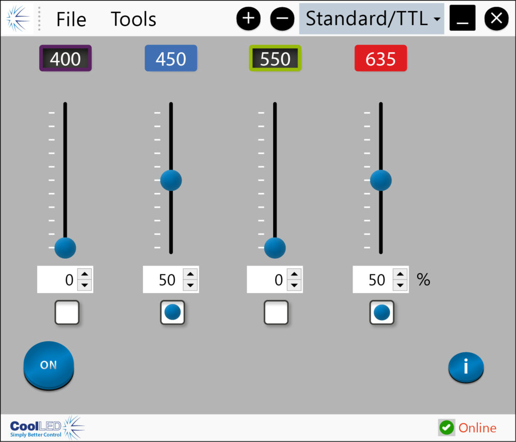
The user-friendly pE-400max LightBridge Graphical User Interface (GUI), provides an additional layer of control, with options including:
- On/off control
- LED selection
- Real time irradiance control
- Sequence Runner
- Save and load pre-sets
- pE-400max start-up settings
Sustainable illumination
The compact pE-400max Illumination System builds on award-winning CoolLED technology, with stable and reliable performance and ultra-low power consumption. The sustainability benefits go beyond energy efficiency, and by removing the need for toxic mercury, the pE-400max is a natural choice for cleaner, greener labs.
Fit to your microscope
The compact pE-400max is compatible with the majority of microscope models, with two light delivery configurations available: direct fit and liquid light guide.
Direct fit variants achieve higher irradiance by directly connecting to the microscope via dedicated microscope adaptors, with a simple once-only adjustment optimising the optical path of the microscope. Not only do these variants avoid extra loss of irradiance as the liquid light guide degrades over time, but they are also more cost-effective. This is because they avoid the need to replace the liquid light guide when it comes to the end of its life.
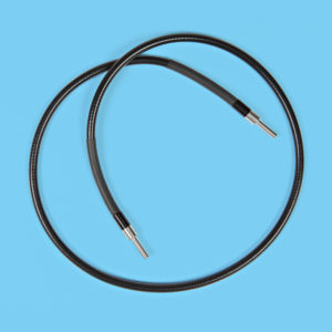
If there is a requirement to keep the source of illumination remote from the microscope, liquid light guide variants are available. These require a fixed 3 mm diameter liquid light guide, and an optional pE-Universal Collimator can be specified in conjunction with a microscope adaptor if required.
Match your DAPI filters
DAPI can be excited at either 365 nm or 400 nm and microscopes typically have a filter set which allows one or other of these wavelengths to be transmitted. To accommodate this, the pE-400max can be specified in one of two waveband configurations to match existing filter sets within the microscope.
A microscope which is populated with a number of single band (“SB”) filter sets will typically have DAPI excitation at 365 nm and a microscope with multi-band (“MB”) filter(s) has excitation at 400 nm. The user can specify the configuration which is appropriate for their microscope.

